Thursday, March 30, 2017

Why Does the "Position Effect" Matter?


What is the Position Effect?

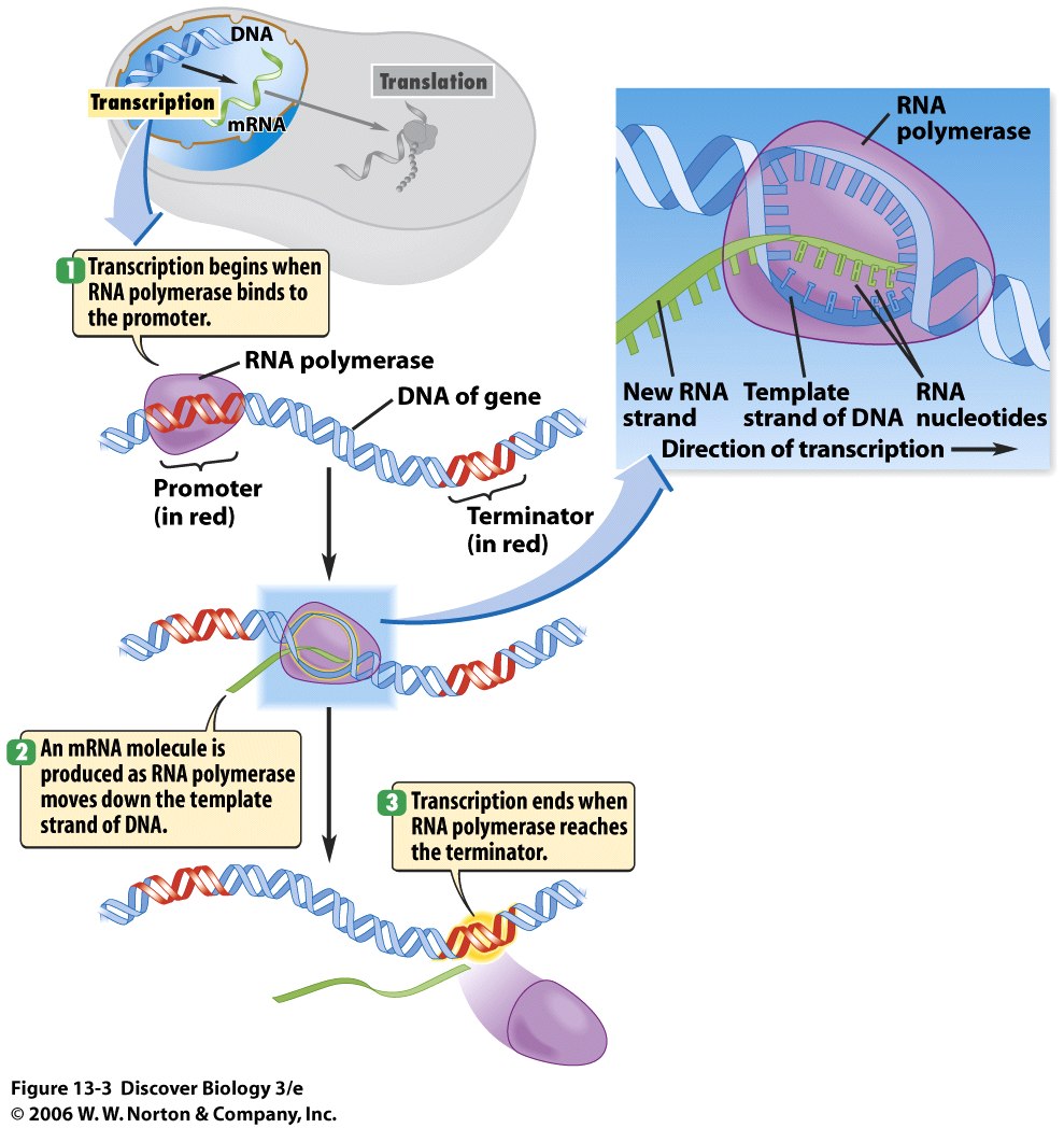
Image result for pictures of transcription
Here is the pathway for transcription.
          There are two different definitions for the position effect that I would like to share in this blog. The first definition is that the position effect is a change in the expression of a gene after a chromosome's position is disrupted. Usually when a chromosome's position or a portion of it is then it's a result of translocation. A translocation occurs when there is a rearrangement of portions of a chromosome that take place between two nonhomologous chromosomes. Like any change in a chromosome, translocation can also occur due to a mutation, crossing over, or even mistakes in meiosis\mitosis depending on the type of cell (somatic or gamete). Not only that but the position effect can describe the variation of how a gene is expressed by identical transgenes. Notably the difference in the gene expression is due to enhancers that regulate other nearby genes. Enhancers are short DNA regions that are bound by proteins called transcription factors that stimulate transcription to occur.
Image result for pictures of translocation

                                                   Here is a picture that displays translocation
          
            What allows these enhancers to stimulate transcription to occur is because of its location near the promoter, which is the region of DNA that forms the site for which transcription can occur. What is so interesting about these enhancers is that they don't have to act upon the transcription site in order to stimulate transcription. Enhancers can instead be bound by activator proteins, which are specialized proteins that coordinate DNA polymerase and transcription factors to transcribe a gene sequence. However I almost forgot to mention that enhancers can be found in the introns, which are the noncoding gene sequences that become separated from a chromosome via RNA splicing, where because of this it doesn't matter if the enhancer is inserted into another chromosome (via translocation) because it will still affect transcription somehow. I haven't been able to find a reason why this still occurs. These enhancers are a really extraordinary topic to continue studying, because there are many enhancers that can be found in the human genome. One example is the enhancer called HACNS1, which researchers today believe had contributed to the evolution of the opposable thumb rotation and the ability to walk on two feet. These enhancers are the key topic that I believe everyone should continue to study, because there is a lot of researcher behind their possible functions in biotechnology.  
Image result for pictures of enhancers interacting with transcription factors
Here is a picture of the interaction between enhancers and the activator proteins.


Monday, March 27, 2017

The Importance of Agroinfiltration to Gene Delivery


What is Agroinfiltration?

                Agroinfiltration is a laboratory technique where scientists inject a suspension of Agrobacterium tumefaciens into a plant host in order to deliver a gene. The delivered gene would be expressed by the Nicotiana benthamiana host, where the plant host can synthesize the target protein(s). During this technique, Agrobacterium tumefaciens is injected into the plant host through either direct injection or vacuum infiltration. The reason why agroinfiltration is valuable to researchers is because many effective recombinant proteins can be produced over a relatively short time period. In the next paragraph, I will discuss the two different avenues to performing this technique.

              By direct injection, what I mean is that a scientist will have to grow the Agrobacterium strain in a liquid culture and suspend the bacteria into a buffer solution. After several hours, the bacteria should be placed in a syringe where the scientist can then inject the bacteria onto the underside of a leaf. Once the bacteria is injected the bacteria will enter the plant through the small openings of the stomata, that are located all over the leaf. When this whole process is finished, the bacteria should be reproducing inside of the plant, until there are enough daughter offspring for the bacteria to highjack the plant machinery to produce the encoded proteins that are held in the bacterial genetic material. Other than that that is pretty much the entire procedure to performing direct injection.

            Now in order to perform vacuum infiltration all you have to do is first place any leave matter, leaf disks, or even the plant itself into a beaker filled with the buffer solution. Wait until the leaves or whatever sample you are using is fully submerged in the solution and place the beaker into a vacuum chamber. Once the leaves are in the chamber turn on the vacuum, allowing the Agrobacterium solution to be forced into the stomata, which is a tiny opening in the plant tissue where gas exchange occurs, and into the mesophyll layer. This will allow the solution to be in contact with almost every plant cell that is found in that layer, which over time will give the cells the necessary genetic information to construct the encoded proteins.  

             How does the bacterial genetic material become processed? When the bacterium Agrobacterium tumefaciens gets injected into the leave, it remains around the intercellular space in order to transfer a combined gene with TI plasmid-derived T-DNA, which is the generic foreign DNA that the bacterium possesses. The bacterium will copy the gene as much as possible so that it can transfer the genes to as many plant cells as possible. The gene of interest will become transiently expressed, which means the genetic material is inserted into a eukaryotic cell in order to produce the target proteins, by all of the infected plant cells. After the genes become expressed, the plant organelles or "machinery" should be utilized to produce the target proteins. Once the proteins are made that is the moment during my laboratory where I can purify the proteins, which I discussed in an earlier blog, and harvest that protein, where in my case I want antibody proteins. Pretty much from there that is all that has to do with agroinfiltration. Believe me I  have discussed the different methods that are used in this entire process, so now I just want everyone to realize that all of the different methods I have discussed; all really come together in my project.  


Friday, March 24, 2017

How Today's Pharmaceutical Companies Expand Gene Delivery


How Lettuce Can Produce Recombinant Proteins (RP's)

         What is a recombinant protein you may ask? A recombinant protein is simply a protein that is made from the combination of several, different genetic material from other living\nonliving organisms. For instance, after reading Dr. Chen's article, Gene Delivery into Plant Cells for Recombinant Protein Production, I learned that the recombinant proteins he and his grad students are attempting to synthesize obtain their genetic material from mammalian, bacteria, plant, and insect cells. These proteins are responsible for the creation of the highly-valued antibodies that hopefully will help prevent viruses such as the Dengue and West Nile virus from replicating. As you can see, it is very important to study further about these proteins because they can benefit the medical world and hopefully bring an end to these disastrous epidemics. The process to producing a recombinant protein is rather simple, but really monotonous in the laboratory. Here is a diagram showing one way how recombinant proteins are made.
Image result for Recombinant Protein Antibody production

           Today's pharmaceutical companies have approved of the use of plants such as Nicotiana benthamiana to produce the recombinant proteins ever since the development of the first plant-derived therapeutic enzyme for Gaucher's disease. Gaucher's disease is a hereditary disease that causes a buildup of fatty tissue around the liver, spleen, and bones. The reason there are a buildup of fatty issue is due to a lack of production of the key fat break-down enzyme called glucocerebrosidase. The buildup of fat around the bones actually weaken the bone tissue and can cause an increase risk for bone fractures due to too much pressure on the bones. After this new treatment, enzyme replacement therapy, was discovered; the transition to plant gene delivery was seen more beneficial than ever. Gene delivery to the transgenic Nicotiana benthamiana plants is not only cheaper in terms of manufacturing costs, but the process is really simple. All I have to do is make sure the target transgene, which is the gene that I extract from another entity and transfer it to a target host to express it, is integrated into the plant genome. Like any other living organism it is at the genome where the genes become expressed and soon produce the proteins that are encoded from those genes' DNA sequences. However, recently a new strategy has been discovered regarding an even quicker way for the recombinant proteins to be produced from the transgenic plants. It turns out that recombinant proteins can be sequenced and produced by the transgenes directly, where the transgene being inserted inside the plant cell can manually use the plant machinery to produce the RP's directly. This process is done only if the 'position effect' is eliminated. I will talk about more about the concept of the 'position effect' in my next blog, because it is a very important concept to understand in my senior project.

           Lettuce is one example of a non-Nicotiana plant host that can produce recombinant proteins, however at a cost. Lettuce is known to produce higher levels of phenolics and alkaloids, which are compounds that can affect purification resins and hence add to production costs. Since there are a higher concentration of these compounds, there are a lot of FDA regulations that lettuce-produced recombinant proteins do not abide to; hence adding to more problems over manufacturing costs. For any con there has to be a pro. When lettuce undergoes the agricultural process called agroinfiltration, which I will discuss more about in my next blog, lettuce is able to produce efficient, abundant recombinant proteins. There is a study where researchers injected deconstructed viral vectors into normal, wild type lettuce hosts in order to observe the expressed pharmaceutical proteins. These researchers used a capsid protein that a combination of Norwalk virus (NVCP) and geminiviral vector genetic material to show that lettuce is efficient in producing recombinant proteins. The tested lettuce sample was able to produce the Norwalk virus like particle as efficiently as a Nicotiana plant host could do as well, where these functioning virus like particles (VLP's) can be used to induce an immune response in lab mice. As you can see they are producing a vaccine just by experimenting with commonplace lettuce that practically every individual can obtain from a farm.  The production of these VLP's are very beneficial to the production of vaccines, since their similar structural characteristics to the real pathogenic viruses can aid in developing immune responses against the pathogens in lab hosts and hopefully in the future for humans as well. Imagine a world where there is a vaccine for every single kind of pathogen. Lastly, since lettuce can be easily grown and organized in controlled acreage environments this adds to another pro to using lettuce as a VLP producer, and hopefully this manageable crop can be further studied to see if it can indeed create a VLP for every virus so that vaccines can be produced.   


Monday, March 20, 2017

Another Day in the Laboratory


The Amazing Application of Gel Electrophoresis


         Last Tuesday in the laboratory I helped made DNA gels using the technique, gel electrophoresis. In order to construct the gels, I needed two key substances, which were agarose and the buffer TAE. Without these key substances, there would be no way for me to be able to extract the DNA sample from a DNA plasmid. A key note I have to address about the plasmid is that we extracted the plasmid from the plant virus, Potato virus X. In this blog I will be talking about my experience performing the entire gel electrophoresis technique, and I will describe how the technique works.

         First off, gel electrophoresis is a laboratory technique used to separate out the DNA fragments of a certain plasmid based off of the DNA's size and charge. In my case, this technique is extremely important, because I have to separate out the junk DNA from the essential or specific DNA strands that is found in the plasmid sample. Usually the plasmid sample appears to be a clear liquid contained in a tiny test tube. Also to point out junk DNA are extra strands of DNA that an organism or nonliving entity such as a virus picks up from either the environment through the process called transformation or from other organisms or viruses through the process called conjugation. The specific DNA strands that I desire encodes for the construction of the antibodies that my whole project is entirely based on. As you can see this technique is very key to my project.   
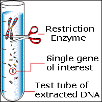
Image result for picture of dna plasmid in a test tube  
Here is an example of a plasmid extract in a test tube
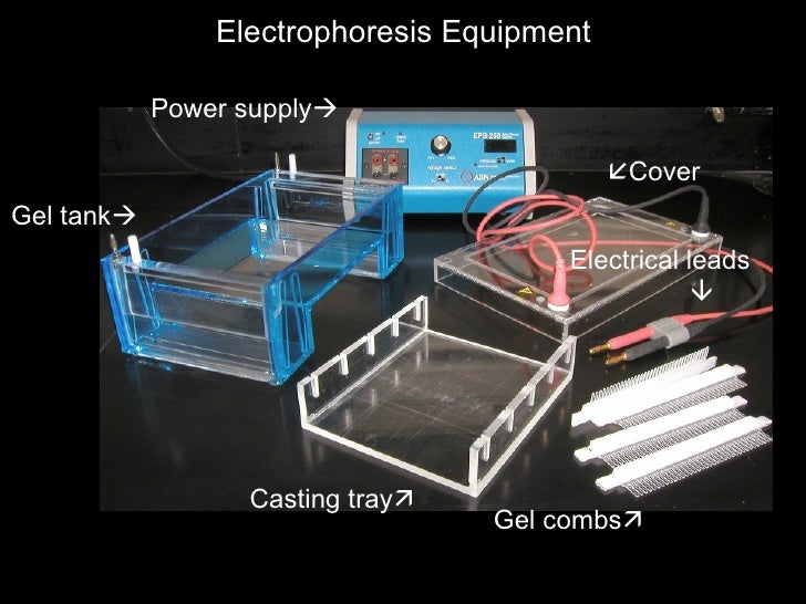
Image result for gel being prepared in gel electrophoresis
Here is a picture of some of the equipment needed to perform gel electrophoresis
               

           The entire process for gel electrophoresis is actually quite simple than most people think. In this experiment, I had to first prepare the gel by using agarose gels to show the DNA fragments according to the length of the individual fragments located on the wells. In my situation, I had to use around 1% of the agarose concentration to perform my gel electrophoresis experiment, because the wells that I had in the lab were really small while the DNA strands that I had were relatively larger than most DNA samples. I then insert the agarose gel into the casting tray and heated it using an electrical current, that was derived from a low voltage power source. Typically it took me around one hour to two hours, because of how low the voltage is in the source. It definelty took too long just for this one step but surely after I completed my gel the whole process I figured the entire process would be longer than I initially presumed. After the gel was heated, I grabbed a comb and placed it at the ends of the tray in order to make well holes on my gel.  After the gel is heated, I had to insert the gel into the gel tank, so that I can finally perform gel electrophoresis. I then inserted the blue TAE buffer fluid onto the gel until the surface of the entire gel is covered, so that the buffer can separate out any DNA and RNA molecules found within the sample plasmid. The key components of TAE that allows for it to perform this function are acetic acid, its Tris bases, and ethylene diamine tetra-acetic acid, where these different acids are responsible for sequestering out the positively charged cations. Remember DNA is typically negatively charged where by removing the cations evidently the DNA molecules are being separated from the RNA molecules. Once the buffer completely covers the gel I had to shift my focus to preparing the DNA from the plasmid sample.

Image result for pictures of a comb being used to separate the wells in gel electrophoresis

Here is the comb "technique" I used.


Preparing the DNA
           This step is by far the easiest step to performing gel electrophoresis, because of how simple it was in the first place. Using a pipette, I inserted a known green dye into the DNA plasmid sample, so that the viscosity or tendency for a substance to resist movement of the DNA increases. This is very important for the entire process, because now the DNA, that will soon be inserted into the wells, cannot float out of the well and also somehow allows the movement of the DNA to be seen on the gel through fluorescent imaging. I then inserted a DNA marker pf known fragment lengths into the first well in order to approximate the size of the other DNA fragments in the other wells. The remaining DNA sample found in my plasmid test tube are then pipetted into the rest of the wells, and finally I am able to close the lid of the electrophoresis tank and play the waiting game. I have to say this part was very simple but really boring because of the long wait for the electrical current being transmitted from the electrodes, that are inserted into the tank.


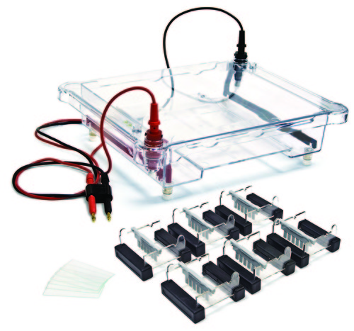
Image result for picture of electrodes being connected to an electrophoresis tank


Here is a picture of the electrodes being assembled onto an electrophoresis tank


How to Separate the DNA Fragments

              In gel electrophoresis, separating out the DNA fragments from the DNA sample is crucial to separating the different DNA strands such as the desired DNA strands that encode the antibodies and the junk DNA strands, that I don't need. Once I have the correct electrodes in their proper positions; I had to wait for the electrical current to be transmitted through each of the DNA wells, so that I can see if the DNA is migrating. Remember DNA is negatively charged so it would make sense for the DNA to be attracted to the positive cations that are surrounding the anode. It was really neat to see how the DNA is migrating from one well to another through monitoring the movement of the loading dye. Like I referenced before in order to monitor the movement of the loading dye; I can see the dye through fluorescence imaging. It was so neat to see the machine that performs this process, because truly it has done a lot of good for today's scientists than ever before. Overall this entire process took me about two days to complete, which basically shows how long it can take to do a simple yet intriguing process such as gel electrophoresis.

Image result for gel fluorescent imagingImage result for pictures of fluorecent imaging in gel electrophoresis
  Gel imaging machine


Friday, March 10, 2017

Understanding the Plant Anatomy is Crucial


Understanding the Plant Anatomy

          Today's lab work was very interesting, because I finally got to work with the Nicotiana benthamiana plants. Frankly, initially I presumed I was going to be able to study the plant production of the antibodies, but instead I was able to work on something more straightforward. I got to feed the plants and "groom" them. I do admit the term "groom" is the wrong diction, but frankly that is what I did today. First, before I started "grooming" and feeding the plants I learned more about the plant anatomy.  For instance, in today's lab work I learned that the test plants produce a lot of structures called suckers. These suckers are what give rise to the formation of a plant's stem. The reason why I had to remove these suckers is to control the height of the plants. Without the suckers the plants wouldn't be able to form a longer stem, hence the plants can't become taller. In my case, removing these suckers was really annoying because there were too many to remove on 60 plants! I swear even though these structures are miniscule, however I had to carefully remove them. Not only are they tightly connected to the plants, but I simply kept on removing the leaves with them. It was so annoying, but finally I got the job done with minimal damage to the plants. Image result for nicotiana benthamiana plants suckers

Here is a picture of a Nicotiana benthamiana plant with its stem extending upwards
Image result for nicotiana benthamiana stemsHere is an example of a sucker.
          After all of that trouble, I finally was able to feed the plants or simply pour a mixed water and fertilizer solution onto each plant. The process was very easy but yet messy at times. I don't remember exactly how many times I got splashed by the water, but I'm telling all of you my shirt was drenched by the time I finished. It was really extraordinary to see how much water we needed to water all of the plants. It took around eight to nine buckets filled with the mixed solution in order to water all of the plants! Those plants definitely receive more than enough water. Frankly, the whole process was really simple but too repetitive. I do have to admit plant biologists and researchers have a lot of work to do other than studying the plants themselves. Hopefully, more people come to realize that studying plants and their structures have more work than they think.  

Monday, March 6, 2017

How Plant-Made Biologics Will be the Future


A New Age in Antibody Production

               After reading the article, Plant-Made Biologics, it is very important to point out that Nicotiana benthamiana plants perform more functions other than synthesize the desired antibodies. These Plant-Made Biologic (PMB) plants are being used today to mass-produce several vaccines that can help combat infectious diseases being transmitted from the Dengue, West Nile, and HIV viruses. In the article, I found out that researchers use the domain III of the West Nile Virus protein envelope as a "vaccine candidate" for the West Nile virus. Somehow the N. benthamiana plants are able to synthesize antigen proteins that can be used to test the structure of the newly developed receptors, that the pathogens possess. If any of you don't realize that in order for a pathogen such as a virus to hook up on the surface protein, the antigen, of a target host cell; the pathogen has to latch its receptor onto the target's antigens. These newly developed antigens are responsible for the development of new vaccines, because whenever a patient is given a vaccine that individual is given a sample of foreign antigens that causes the individual's immune system to develop immunity by producing antibodies against those foreign antigens. Not only this but the plant-derived domain III somehow are able to resist denaturation, allowing the protein envelope's structure to not change, which allows the function of the envelope to remain intact. However a problem I have read from the article to this is that the vaccines produced from the domain III protein envelope haven't been able to work in humans just yet. So far the plant-made vaccines are being administered in mice.


What It's Like to Work in a Microbiology Laboratory?


              Ever Wonder What It's Like to be in a Laboratory?


As of Monday last week, I finally started my research in the laboratory. I have observed how the grad students at the ASU Biodesign Institute extract the necessary antibody proteins from the Nicotiana benthamiana plants and it is a very simple process. Simple laboratory techniques such as centrifugation, and filtration are used to breakdown the targeted protein from all of the biota, the potato X virus and the lab plants.

The first step I worked on in the laboratory was placing the fully grown plants in a blender in order to mash up all of the plant material into a droopy, green liquid paste substance. The reason why the grad students did this step was to acquire the necessary protein that has been stored in the plant material, which would be used to synthesize the antibodies. After all of the green "paste" is mashed up in the blender, it was neat to see that a simple everyday process called filtration is used. Simply the grad students just grabbed some filter sheets and placed the sheets onto four plastic, homogenous bottles. Filtering out the plant material was a very straightforward process in my opinion. The process is actually quite similar to filtering out coffee, when someone wants to have some tasty coffee in the morning. After all of the pure plant material is filtered and placed into four capped bottles, the grad students simply placed all four bottles into a plastic box and inserted ice to cover the sides of the bottles. It's pretty much the same when you place ice in a cooler in order to cool your refreshments. This step is the most essential one in the overall task, because like any protein there is an optimal temperature to which the protein needs to be in, so that the protein doesn't denature or break down in terms of structure. After 5-10 min the grad students grabbed the bottles out of the ice box and placed them in the centrifuge machine. The centrifuge machine we used was enormous. It looked almost like the size of a dryer! We set the four bottles inside the centrifuge machine and set the rotation rate of the machine to 1400 rev\min so that the protein found in the plant "paste" is layered and separated from the rest of the plant biomaterial. Thanks to all of these techniques I was able to observe how any plant protein can be separated from the plant residue, which allows me to further appreciate plants more.