Tuesday, April 25, 2017

Results From the Laboratory


To Think How Much Work Was Achieved


                   Last week while working in the laboratory; I received news about the data collected for the amount of viable antibodies that were produced from the infiltrated plants. It turned out that for about each gram of leave material that was infiltrated around 350 mg of antibodies were produced! So far as I know the mass of the leaves is uncertain because in the laboratory I am dealing with several different sizes of leaves, making it difficult to figure out exactly an average mass for these plants' leaves. Typically the mass of the leaves I have dealt with can be around 5 to 7g of leave matter, but again the amount of leaf matter that is still available after infiltration can change.This number is super tremendous because thinking about it instead of injecting a common vaccination into a patient such as say giving the patient a subunit vaccination, which is injecting the pathogenic antigen into a patient's body, doctors can turn to injecting human-like antibodies into the patient. Think about it imagine injecting several hundreds of mg of antibodies into a host, where those antibodies can be used to combat the replication of a pathogen found inside a host without the need for the host's immune system to respond to the infection directly. Typically in most individuals the rate at which a human immune system produces its peak amount of antibodies that can respond to a specific strain of a virus is typically two weeks. Two weeks is a long time and by that time the infection could have gotten worse as the  virus mutates inside the human immune system, causing the virus' shape and antigen structure to change with it. Not only that but the usage of antibiotics to destroy the virus wouldn't work too because antibiotics can only target against bacteria and not viruses. The implementation of plant-based biological systems has greatly posed a better future for vaccination alternatives.
Image result for picture of a primary immune response


                  Although I have brought up some pros to the implementation of the plant-produced antibodies, there are still cons to this alternative that have to be addressed in the given future in order for this system to be efficient for a greater tested population. One obvious con that I hope everyone reading this asks to themselves is how can there be enough antibodies produced to supply for a larger population. Typically the production of these antibodies can take around 2 to 4 months, because the researchers have to grow the Nicotiana benthamiana plants and control their growth. In that case, there has to be thousands of plants to be produced and grown in the right conditions in order for there to be enough antibodies to be produced from these lab organisms. Not only the amount of time it takes for the plants to be produced but the decision to continue using Agrobacterium, PVX (Potato virus X), and TMV (Tobacco Mosaic virus) comes into play, because there has to be enough of these organisms to be grown to transmit the correct gene sequences for the plants to use to produce the antibodiesAlso, there is a problem with using antibodies to defend a human body, because the antibodies are specific to one type of viral structure (active site) and so if the virus was to mutate then there is no way for the introduced antibodies to even work. Typically when making the antibodies there has to be enough buffer solution such as MES to be able to help stimulate growth of the bacteria, so that task right there can be a problem. The timing of this entire process and the issue that adaptive immunity isn't stimulated in a human immune system can be difficult potentials to the use of plant-made antibodies. It's better for immunity to be given to the human hosts instead of giving the patient antibodies by injecting a vaccination, but there is always an issue with viruses being able to change their structure due to mutations. This ability that viruses have is something that has to be addressed in order for any of these two mechanisms to work efficiently for the future populations that are exposed to viruses such as Dengue.
Image result for picture of adpative immunity
Here is a picture of the two types of adaptive immunity and the type of immune cells utilized in each type


Monday, April 24, 2017

Glycosylation and its Applications to the Transgenic Plants


Glycosylation and the Transgenic Plants


                 What is glycosylation, you may ask? Is it a process, a part of a plant, or even an artificial machine? No, but rather it's a mechanism that is performed by plants that is very crucial to understanding my project. Glycosylation is an enzymatic, site-specific process that takes place around the endoplasmic reticulum where a carbohydrate molecule is added to a target protein. In this sense, the carbohydrate helps to stabilize a protein's structure, allowing the protein to fold correctly. Another purpose for glycosylation is that it helps immune cells to be able to recognize host cells by developing sugar-binding proteins called lectins. These lectin proteins are what allow the immune cells to recognize other host cells by recognizing specific carbohydrate molecules, that are found on the surface of all host cells. As you can see clearly, this property is very important for a healthy immune to function property and not end up becoming an autoimmune disease.There are five different classes of glycosylation and they include: N-linked glycosylation, O-linked glycosylation, phosphor-linked glycosylation, c-linked glycosylation, and glypiation. For the development of the monoclonal antibodies at the Biodesign Institute, the researchers utilize N-linked glycosylation to change the structure of the Fab region of the antibodies. The Fab region of an antibody is the portion of the antibody that locks or hooks onto a specific cell surface for which in the case of antibodies is what allows the antibodies to opsonize a particular pathogen.     
Image result for picture of the fc region of an antibody
Here are the three regions that make up an antibody.

             Hence, as you can see by changing the structure of the antibodies the researchers at the Biodesign Institute are altering the role of these proteins so that they can opsonize the specific Dengue virus. If I haven't mentioned it before, what I mean by opsonization is that it's an immune defense process where antibodies specific to a particular virus can surround the entire virus surface and hook onto the virus' surface receptors, keeping the virus from being able to infect a host cell and replicating. This ability to alter the structure of an antibody is very crucial to my project and is key to unlocking a new alternative to treating pathogens such as the Dengue virus.

               At the Biodesign Institute, researchers modify the N-glycosylation process that normally occurs in the Nicotiana benthamiana plants. Usually N-glycosylation requires a special lipid called dolichol phosphate to help attach a nitrogen molecule from asparagine with the glycan, a regular carbohydrate molecule found in plants. N-glycosylation is very unique because normally this enzymatic process occurs in the lumen, which is a network of membrane tubules and vesicles that are responsible for the production of hormones, of the endoplasmic reticulum in eukaryotes. This process is responsible for the folding of many eukaryotic glycoproteins and the cell-extracellular matrix attachment. However at the Biodesign Institute, N-glycosylation to enable proteins in the lab plants to produce the desired antibodies. Normally, since the plants are receiving genetic material from several other bacterial\virus species\strains the plant's immune molecules would attack the foreign cells\viruses. Due to our work at the Biodesign Institute, we were able to modify the glycoproteins' structure found on the surfaces of the introduced bacterial cells and viruses in order to mask their foreign identity from the plant and mammalian immune systems. Somehow by modifying N-glycosylation researchers are able to change the structure of the antibodies, that would be produced from the plants, so that these antibodies appear mammalian-like. As you can see these antibodies like the bacterial cells\virions will be able to float around a host's immune system and perform its duty without being detected by the host's immune system and being destroyed. Overall, the main job researchers want from N-glycosylation is for genetically modified antibodies to be synthesized that can be undetected from both the plants' and mammalian' immune systems.

                 That is the end to this blog but stay tune to news about the data collected from the antibody quantity produced from the Nicotiana benthamiana plants.   


Wednesday, April 12, 2017

Heat is the Answer to Cleaning


How to do-Autoclave?

                       Autoclave is a laboratory cleaning method where I have to place contaminated lab equipment into this oven-like machine to kill any bacteria, that would reside on the equipment. The lab equipment may include: beakers, flasks, graduated cylinders, lab bottles, and the lab bottles' caps. Before I put the equipment into the oven, I had to clean out all of the dishes using dish water and bleach so that I can kill as many bacteria as possible before the rest of the bacteria and their endotoxins are destroyed in the oven. Also, remember since I am working with glass beakers, flasks, and plastic bottles I will have to put aluminum foil to cover the ends of the equipment, so that the bottles, beakers, and flasks don't get damaged from the heat. Simply just like any household oven remember to wear oven mitts, because inside the autoclave machine is very hot. Don't be the person who gets burned from accidentally touching the inside of the machine. Believe me the machine is very hot and you can sustain a second degree burn from this machine! After I placed all of the lab equipment on plastic trays, I then place the trays into the machine and set the timer for about 2 hours. Lastly, once the two hours were up all I have to do is get rid of the aluminum foil and put all of the lab equipment away in their designated places. Pretty much that is all I did when performing this method. It is pretty simple enough, but remember to wear oven mitts, because I have already been burned from this machine and it sure hurt a lot!

Image result for pictures of an autoclave oven
Here is a picture of the inside of an Autoclave machine
Image result for pictures of using an autoclave machine


How Important are Pellets in a Laboratory?


How Are Bacterial Pellets Made?

               On Friday, for the first time I learned how to grow bacterial pellets. The entire process was pretty repetitive and time-consuming, but overall the whole agenda was interesting. First off, I want to clarify what is a bacterial pellet and why we, researchers at the Biodesign Institute, need to grow them.

                A bacterial pellet is a clump of dense bacterial cells that grow together in a laboratory bottle due to a centripetal force. What produces the centripetal force, you may ask? Well, in the laboratory we use a centrifuge to produce this force as we spin down two or more balanced bacterial media bottles; in order to separate out the bacteria cells from the liquid media. The most important rule I learned about using a centrifuge is to make sure the lab bottles are balanced. By balanced I mean that both bottles have the same mass amount by using a common mass scale. In my case, whenever I needed to add more media solution into one of the bottles; the trick is to just add water into the less massed bottle, until the mass of the bottle is about the same as the other bottle. The reason why the bottles have to be balanced is so that the rotor in the centrifuge doesn't get damaged by spinning so fast where the unbalanced side of the cup holders doesn't teeter and totter, causing the rotor to break. In our case, we spun the bacteria at around 5000 rpm, which is very fast if any you get the chance to see it. It took around 10 to 15 minutes for us to spin the bottles down, but all the work was worth it since the bacterial pellet grew. See picture:
Image result for pictures of growing bacterial pellet in a laboratory bottle
The brown patch in the picture is the bacterial pellet.
Notice how compact the pellet appears, where this tells me how the bacterial cells are clumping together due to centripetal force.
So far I have only talked about how the bacterial pellets grew, but now I still need to answer one of my questions about why we need to use them? When we grow bacteria particularly the Agrobacterium strain, we are focusing on bunching up all of the bacterial cells together so that we can have a huge concentration of these cells to infiltrate. Hence, the bacterial cells will clump together to form the pellet, as shown in the picture. The more concentrated bacterial cells we have then the more concentrated media solution we can produce to which the gene of interest, the gene that codes for the antibodies, can be encoded in the plants' tissue. In order to grow the media solution, I had to put the bacterial cells in a MES buffer solution, so that the bacteria can grow in the solution and be stimulated to transfer genetic material to each other. As the bacteria are growing in the solution, I have to then pour a little sample of the solution into a cuvette, which is a small container that fits into a spectrometer, so that I can place the cuvette into the spectrometer to measure out the optical density of the bacteria cells in the sample.  What the spectrometer does in order to give me the optical density of the sample is by passing a 600 nm wavelength light through the cuvette, where any light that was blocked by the cells themselves would be determined from the amount of light that was able to pass through the cuvette, which is measured out by the machine. The density value will then be used to calculate out an estimate of how much bacterial cells there could be in these large 500mL flasks, that contain the bacterial cell "waste" solution left out from the pellet.

Here is a picture of a lab bottle filled with concentrated bacterial cells after using the vortex
Whenever I use the centrifuge, the liquid that is still in the lab bottles where the pellet forms at the bottom is disposed into labeled bacterial strain flasks. The liquid may still has some bacterial cells in the solution, but most often then not it will be the MES buffer solution that I mentioned. I keep pouring in more solution into each specific lab bottle, until the pellet has formed completely. After that I can use a vortex spin machine to grind out the pellets to dilute the bacterial cells into a clear white liquid. That white liquid will be added to another solution of MES where then it can be used for infiltration. Other than that that is pretty much all the skills you will all need to know in order to make a bacterial pellet.
Image result for vortex machine
Here is a picture of a vortex machine.


Thursday, April 6, 2017

How it's Like to Perform Agroinfiltration?


Agroinfiltration Firsthand

              In an earlier post, I have talked about what is agroinfiltration and how it is used in today's pharmaceutical companies to develop antibodies. After yesterday's lab day, I have finally performed agroinfiltration for the first time. Frankly, the entire process wasn't really difficult to perform but rather repetitive. All of the supplies I needed were a needle, a 10 mL syringe, several trays holding the plants, and a 100mL beaker filled with some medium solution. The medium solution looked clear and viscous, and luckily the solution wasn't harmful to the skin, because even with wearing laboratory gloves some media spilled on my skin. The quick solution to this occasion was to wash my skin with water, so like I said before the solution is safe. In the medium solution stores bundles of key nutrients that the plants need to grow and the Agrobacterium that hopefully will deliver the needed instructions for the plants to produce the desired antibodies. I used the syringe to pull out the solution from the beaker and with a needle I poked a small incision on the leaves' undersides. As you can see from the picture, next I placed my index finger on the top portion of the leaf and placed my syringe on the incision that I made from the needle, so that none of the medium flows out of the leaf. This process continues repeatedly until the leaf is completely filled with the medium, where as you notice this will end when the dark patch is spread all around the entire leaf.

Here is a picture of a scientist performing agroinfiltration by injecting medium inside the leaf.
Notice how the solution spreads throughout the leaf as shown by the dark patch.

              Like I said in my previous post agroinfiltration is an elaborate technique to  "convince" a plant's machinery to produce antibodies whose codons were delivered from another bacterial species. This technique is very simple yet can be missing when you are doing this for the very first time. Trust me I struggled a lot with making a small incision with the needle, because I would place too much pressure and end up poking a hole through the leaf. Other than that this method is very simple but can take a long time depending on how many plants you have to infiltrate. In my session, I had around 60 plants to infiltrate and that took about 2 hours for me to do with a lab partner, so this process is pretty long but repetitive. If anyone decides to perform this procedure, just remember to bring some headphones because this whole process can take a long time.


How VLP's Are Key to Future Vaccinations


VLP's Are the Key to Vaccinations

              Imagine a world where immunogenicity is offered to every individual all at a price of a vaccination shot. Thousands to millions of lives could be saved from deadly, epidemic diseases just by taking a single shot. Is it worth it? This is the world that I want to pursue and hopefully with the help of Nicotiana benthamiana  plants it will be possible. In this article that I have read, which is called Plant-derived Virus-like Particles as Vaccines, there are specialized particles called virus-like particles (VLP's) that are used to help the host immune system develop immunity against a certain surface protein or antigen. However, something to point out VLP's only help develop immunity in a host only against pathogenic viruses and not bacteria. These VLP's derived their unique structure from viral antigens that mimic the general structure of the real viruses, however these VLP's lack the viral genome. Due to a high presentation of viral antigens on a natural virus' surface, VLP's are able to copy these antigens and mimic the antigens on their surface. This result demonstrates that VLP's are valuable in vaccinations for humans, because our immune system just has to encounter these antigens for a first time, before our immune system develops antibodies that are best suited to those specific antigens' structure. The major problem to VLP's is the amount of production costs, but in this article a solution has been made.

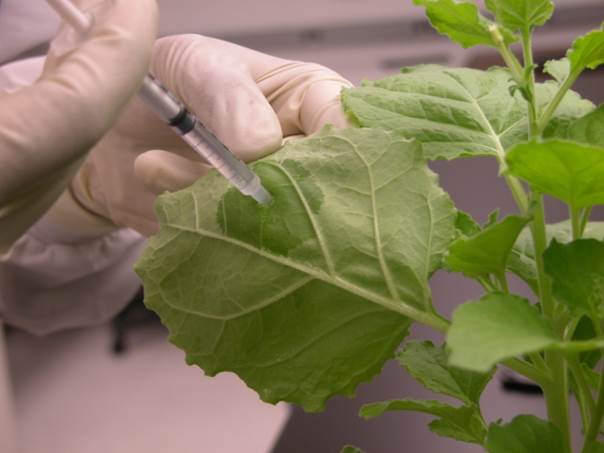
              The lab plants, Nicotiana benthamiana, are a cheaper alternative to producing these VLP's, because of the low production costs, low maintenance costs, and high scalability factors that are offered by these plants. The production of VLP's in plants is a quicker process than other mechanism because the transgenic plants that I am working with grow quickly when placed in the right laboratory conditions: high humidity, water tank, fertilizer, and a healthy soil amount. It takes around two weeks for the plants to grow to the max controlled height that we want the plants to be up to, so that shows to tell you that these plants grow really quickly. What is so remarkable about these plants is that according to a theory about vaccine transportation; plants that hold the VLP's can be ingested in edible plant parts such as the leaves in order to transmit the VLP's particles orally to a host. However, this is only a theory and has been only tested in lab mice. Also, to point out there are several concerns over this delivery system such as possible denaturation occurring to the proteins that make up the antibodies due to the low pH in the human digestion system, poor recognition of the vaccine at mucosal immune effector sites, and antigenic tolerance. As you can see the biggest hurdle to using plants as an antibody production site is the delivery method of the vaccination to a host. In the article, the author mentioned that the most reasonable delivery method would be direct injection using a syringe needle, which is the most common alternative for any vaccination. In the next paragraph, I will be discussing how the VLP's develop immunity for hosts like humans against the actual deadly pathogens.

                  The way the VLP's are structured is the main reason why these simple particles can help hosts develop immunity against the wild type viruses. Since VLP's are particulate it allows them to induce T-cell mediated immune responses due to their interaction with the antigen presenting cells (APC) found in the host's immune system. Remember T cells are specialized immune cells that work to destroy a foreign cell by signaling the infected cells to undergo apoptosis, which means the cell self-destructs and dies. This action is vital to a host's defenses, because of how effective this process can be directed against pathogens. In order for the VLP's to induce the T cell activation they have to  mimic the process of a natural infection, where the antigens presented by the VLP's should trigger a cascade immune event where the host's immune system attacks the VLP's. As the immune system destroys the VLP's, since the VLP's have no mechanism to infect the host's cells, the B cells should develop "memory" against the specific antigen structures that were presented by the VLP's. In turn, the host's immune system would develop immunity against the actual pathogenic virus, since the immune cells can quickly develop an immune response against the pathogen quicker. Another reason that allows the VLP's to develop immunity in a host is the presence of epitopes on the cell surface. Epitopes are a specific region on an antigen that is recognized by the host's immune system and to which where an antibody binds to. The presence of thousands of epitopes on a single VLP aids to the processing and presentation of APC's.  What else is so interesting about these VLP's is that some of them are so small that they can diffuse to the lymph nodes, allowing the VLP's to be presented efficiently by B and T cells. That is all that is to it to VLP's and how they help develop immunity in hosts. These particles are very efficient at their job and hopefully become more available to be used in all future vaccinations.