The Amazing Application of Gel Electrophoresis
Last Tuesday in the laboratory I helped made DNA gels using the technique, gel electrophoresis. In order to construct the gels, I needed two key substances, which were agarose and the buffer TAE. Without these key substances, there would be no way for me to be able to extract the DNA sample from a DNA plasmid. A key note I have to address about the plasmid is that we extracted the plasmid from the plant virus, Potato virus X. In this blog I will be talking about my experience performing the entire gel electrophoresis technique, and I will describe how the technique works.
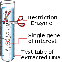
First off, gel electrophoresis is a laboratory technique used to separate out the DNA fragments of a certain plasmid based off of the DNA's size and charge. In my case, this technique is extremely important, because I have to separate out the junk DNA from the essential or specific DNA strands that is found in the plasmid sample. Usually the plasmid sample appears to be a clear liquid contained in a tiny test tube. Also to point out junk DNA are extra strands of DNA that an organism or nonliving entity such as a virus picks up from either the environment through the process called transformation or from other organisms or viruses through the process called conjugation. The specific DNA strands that I desire encodes for the construction of the antibodies that my whole project is entirely based on. As you can see this technique is very key to my project.


First off, gel electrophoresis is a laboratory technique used to separate out the DNA fragments of a certain plasmid based off of the DNA's size and charge. In my case, this technique is extremely important, because I have to separate out the junk DNA from the essential or specific DNA strands that is found in the plasmid sample. Usually the plasmid sample appears to be a clear liquid contained in a tiny test tube. Also to point out junk DNA are extra strands of DNA that an organism or nonliving entity such as a virus picks up from either the environment through the process called transformation or from other organisms or viruses through the process called conjugation. The specific DNA strands that I desire encodes for the construction of the antibodies that my whole project is entirely based on. As you can see this technique is very key to my project.

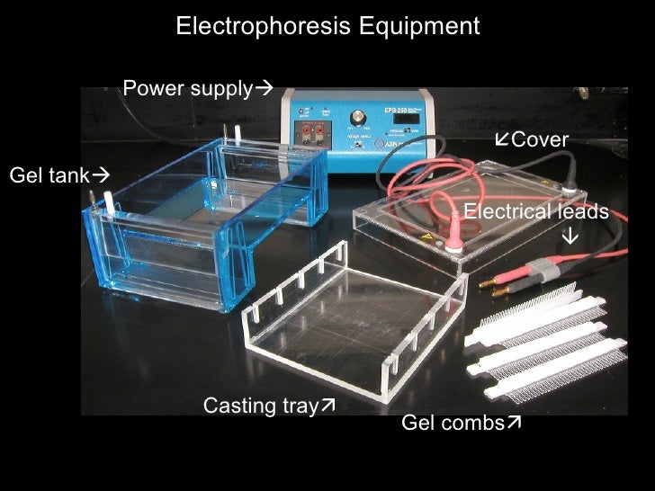
Here is a picture of some of the equipment needed to perform gel electrophoresis
The entire process for gel electrophoresis is actually quite simple than most people think. In this experiment, I had to first prepare the gel by using agarose gels to show the DNA fragments according to the length of the individual fragments located on the wells. In my situation, I had to use around 1% of the agarose concentration to perform my gel electrophoresis experiment, because the wells that I had in the lab were really small while the DNA strands that I had were relatively larger than most DNA samples. I then insert the agarose gel into the casting tray and heated it using an electrical current, that was derived from a low voltage power source. Typically it took me around one hour to two hours, because of how low the voltage is in the source. It definelty took too long just for this one step but surely after I completed my gel the whole process I figured the entire process would be longer than I initially presumed. After the gel was heated, I grabbed a comb and placed it at the ends of the tray in order to make well holes on my gel. After the gel is heated, I had to insert the gel into the gel tank, so that I can finally perform gel electrophoresis. I then inserted the blue TAE buffer fluid onto the gel until the surface of the entire gel is covered, so that the buffer can separate out any DNA and RNA molecules found within the sample plasmid. The key components of TAE that allows for it to perform this function are acetic acid, its Tris bases, and ethylene diamine tetra-acetic acid, where these different acids are responsible for sequestering out the positively charged cations. Remember DNA is typically negatively charged where by removing the cations evidently the DNA molecules are being separated from the RNA molecules. Once the buffer completely covers the gel I had to shift my focus to preparing the DNA from the plasmid sample.

Here is the comb "technique" I used.
Preparing the DNA
This step is by far the easiest step to performing gel electrophoresis, because of how simple it was in the first place. Using a pipette, I inserted a known green dye into the DNA plasmid sample, so that the viscosity or tendency for a substance to resist movement of the DNA increases. This is very important for the entire process, because now the DNA, that will soon be inserted into the wells, cannot float out of the well and also somehow allows the movement of the DNA to be seen on the gel through fluorescent imaging. I then inserted a DNA marker pf known fragment lengths into the first well in order to approximate the size of the other DNA fragments in the other wells. The remaining DNA sample found in my plasmid test tube are then pipetted into the rest of the wells, and finally I am able to close the lid of the electrophoresis tank and play the waiting game. I have to say this part was very simple but really boring because of the long wait for the electrical current being transmitted from the electrodes, that are inserted into the tank.
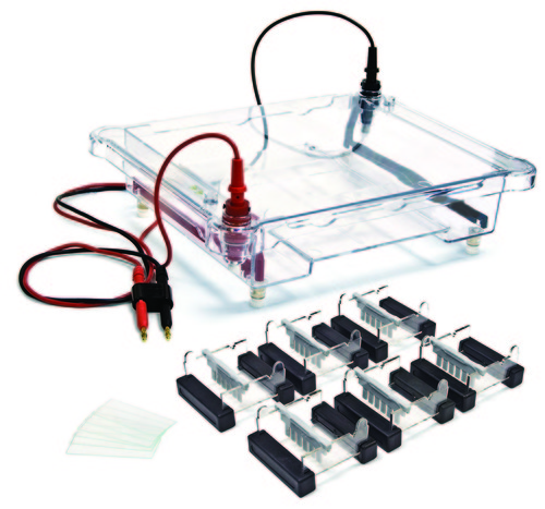
Here is a picture of the electrodes being assembled onto an electrophoresis tank
How to Separate the DNA Fragments
In gel electrophoresis, separating out the DNA fragments from the DNA sample is crucial to separating the different DNA strands such as the desired DNA strands that encode the antibodies and the junk DNA strands, that I don't need. Once I have the correct electrodes in their proper positions; I had to wait for the electrical current to be transmitted through each of the DNA wells, so that I can see if the DNA is migrating. Remember DNA is negatively charged so it would make sense for the DNA to be attracted to the positive cations that are surrounding the anode. It was really neat to see how the DNA is migrating from one well to another through monitoring the movement of the loading dye. Like I referenced before in order to monitor the movement of the loading dye; I can see the dye through fluorescence imaging. It was so neat to see the machine that performs this process, because truly it has done a lot of good for today's scientists than ever before. Overall this entire process took me about two days to complete, which basically shows how long it can take to do a simple yet intriguing process such as gel electrophoresis.
Gel imaging machine


Hi Armando,
ReplyDeleteIt's really cool that you were able to perform a gel electrophoresis! It was only a few weeks ago that we did an online lab and now you get to do the experiment in real life. How many times did you replicate the potato virus X this time around?